 |
Startup/Licensable
Technology: |
|
Startup/Licensable
Technology: |
|
How do I license this technology? Gallery Press Coverage
"Transdermal Photopolymerization for Minimally Invasive Implantation", Proc. Nat. Acad. Sci., USA "Transdermal Photopolymerization of PEO-Based Injectable Hydrogels for Tissue Engineering Cartilage", Plastic and Reconstructive Surgery "Synthesis and Characterization of Photocrosslinked Polymers Based on Poly(L-lactic acid-co-L-aspartic acid)", Macromolecules "Cogelation of Hydrolyzable Cross-Liners and Poly(ethylene oxide) Dimethacrylate and Their Use as Controlled Release Vehicles", Intelligent Materials for Controlled Release, ACS Symposium "Photoencapsulation of chondrocytes in Poly(ethylene oxide)-based Semi Interpenetrating Networks", Journal of Biomedical Materials Research, 51(2):164-71, 2000 "Controlled Released IGF-I and TGF-b on Bovine Chondrocytes Encapsulated in a Photopolymerizing Hydrogel", accepted to the Journal of Orthopedic Research. "Transtissue Photopolymerization of Poly(Vinyl Alcohol) Hydrogels" The Wiley Polymer Networks Group Review Series, vol. 2, B.T. Stokke (ed.), in press "A Tissue-Engineered Conduit for Facial Nerve Repair", Arch Otolaryngol Head Neck Surg, 124(10):1081-6, 1998 "Transdermally Photopolymerized Adhesive for Seroma Prevention", Plastic and Reconstructive Surgery, 103:531-535, 1999 V. Ting, CD Sims, LE Brecht, JG Kasabian, AK Connelly, J.Elisseeff, GK Gittes, MT Longaker, "In vitro prefabrication of human cartilage shapes using fibrin glue and human chondrocytes" Ann Plast Surg, 40(4):413-420, 1998 Selected Abstracts Transdermal Photopolymerization to Create an Injectable Tissue Engineered Cartilage", World Congress on Medical Physics and Biomedical Engineering, Nice, France, 1997 Interactions of light and skin for optimization of transdermal photopolymerization", 44th Annual ASAIO Meeting, Spring 1998 Transdermal Photopolymerization for Cartilage Tissue Engineering", Biomedical Engineering Society Annual Meeting, 1998 Meniscal Tissue Engineered by Suncutaneous Implantation of Fibrochondrocytes on Polymer Develops Biomechanical Compressive Properties Similar to Normal Meniscus", Orthopedic Research Society Meeting, Spring, 1997 Scaffold Materials to Engineer Cartilage", Biomedical Engineering Society Annual Meeting, 1998 Cationic Polymerization of Styrene in the Presence of Ethers and Sulfides", Polymer Preprints, 35(1):464, 1994
|
Introduction:
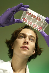
About ten years ago, doctors in Sweden began to surgically remove samples of healthy articular cartilage from the knees of patients with cartilage defects and use it to grow new cartilage in tissue culture. Once they had enough material, they went back into the patient’s knees, removed the damaged cartilage, and inserted the test tube cells, called chondrocytes, in its place. The results were promising. Today, there are three main types of transplants:
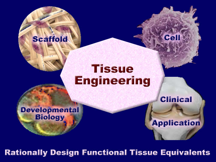
Tissue engineers today utilize a variety of approaches to regenerate tissues. These approaches can be broadly characterized into three major groups. First, biomaterials, without additional cells, are used to convey biological signals to surrounding tissues to recruit cells and promote inherent regeneration. Second, cells alone may be used, without a biomaterial, to form tissues. Finally, cells may be used with a biomaterial scaffold that acts as a framework for developing tissues. The technology focuses on the latter technique, developing and using biomaterial scaffolds for tissue engineering. The development of an effective cartilage regeneration system and future improvements rely on the merger of skills and knowledge as shown below:
The Elisseeff tissue engineering laboratory has applied rigorous basic science and engineering principles to the development and understanding of tissue engineering systems. They
Therapeutic Focus: The Hopkins technology leverages the expertise of basic and clinical medicine, biomaterials, and engineering to provide an improved product with the following properties:
Market Opportunity: Over 15 million people worldwide suffer form knee-joint failure each year due to the breakdown of surrounding cartilage in the joint and the inability of this cartilage to repair itself through the natural regenerative processes of healing in the body. About 200,00 total knee replacements are performed each year at an average cost of $25,000 each, in an attempt to treat this problem. According to industry estimates, more than 225,000 people worldwide underwent arthroscopic meniscal repair in 2002. The number of procedures is expected to have a compound annual growth rate of nearly 5% through 2007, according to a major company in the field. Chondrocyte transplantation is a relatively new procedure and is growing rapidly, in part due to improvements in cell harvesting and in vitro cell expansion. According to one industry player, more than 2,100 people in the United States have had cartilage cells harvested for grafting and implantation in hopes that these cultured cells will relieve the persistent pain and grinding in their knees. The current total cost of the procedure is about $30,000, with the lab cultivation procedure costing $10,000 and the hospital and surgical fees up around $20,000. The primary markets for the Hopkins technology include North America, Europe, and some countries in Asia, such as Japan, Korea, and Singapore.
1998
Comparison of the European Knee Implant Market by Country Clinical Competitor Analysis: There are a number of competitors with products in the marketplace and under development. The distinguishing factors for market success will include efficacy, ease of use, cost, and long term benefits. The Hopkins technology is being optimized not only at the biomaterial's level, but also at the cellular level. Following are some of the major players in the field: Genzyme Tissue Repair's Carticel® and its second generation products represent the market leader at present. Smith & Nephew has introduced a new implant that applies the latest advances in medical technology to an established repair technique in the field of arthroscopic surgery. Called FasT-Fix AB Meniscal Repair System, the system was first launched in April 2001. OsteoBiologics, Inc. is also focused on the research, development and manufacturing of bioabsorbable tissue-engineered scaffolds and instrumentation to identify and repair musculoskeletal tissue. The autologous cartilage transplant CaReS®- Cartilage Repair System - offered by ARS ARTHRO AG® is a 3D mechanically stable transplant based on patient specific autologous cartilage cells and a unique patent pending collagen matrix. BioTissue Technologies has developed the Autologous Chondrocyte Graft BioSeed®-C for the treatment of defective joint cartilage. IsoTis is developing tissue-engineered products, including skin, bone and cartilage for use in clinical settings. Isto Technologies is involved in the development of engineered tissues and chemical compounds for the repair and regeneration of human tissue. Orquest is involved in the development of orthobiologic products for bone and cartilage regeneration, using combined technology from tissue biology and biomaterial science. Osiris Therapeutics, Inc. is a researcher and developer of regenerative human stem cell technologies for eventual use in therapeutic products. OsteoBiologics is a developer and manufacturer of bioabsorbable tissue-engineering scaffolds (IMMIX) for the repair and replacement of musculoskeletal tissues. ReGen Biologics designs, develops and manufactures minimally invasive implants and medical devices for the repair and regeneration of damaged or degenerating cartilage in the knee. There are other therapies such as Synvisc's injectable rooster-derived extracellular proteins and also nonbiological implants, such as Sulzer Orthopedics' UniSpacer, designed to provide reduced friction between the joints. The Development Team Jennifer Elisseeff Principal Investigator jhe@bme.jhu.edu
|
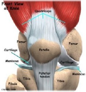
The Technology: The knee joint is very complex. It is composed of four bones, the ligaments that hold them together, the articular and meniscal cartilage that protects the ends, and the surrounding bursa.
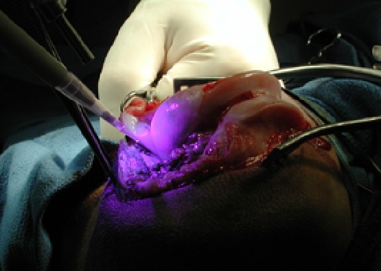
The Hopkins cartilage regeneration technology encompasses a number of separate components to replace damaged cartilage in the knee or in other joints or locations. The technology provides a convenient and effective photopolymerization of a hydrogel that contains cells and growth factors necessary for the growth and integration of new cartilage in a damaged tissue. While cartilage is often considered a simple tissue with chondrocytes sparsely distributed throughout an extracellular matrix of type II collagen and aggrecan, it is a complex, heterogenous tissue. On the cellular level chondrocytes are organized into superficial, proliferating, prehypertrophic and hypertrophic chondrocytes. These different types of chondrocytes have different cell morphology and gene expression. The extracellular matrix around the chondrocytes is also highly organized and changes depending on the where it is located. The cellular and extracellular matrix organization is controlled by mechanical and biological signals during development and after. There is a large wealth of molecular and cell biology that has been applied to cartilage tissue engineering. This technology uses hydrogels for tissue engineering.
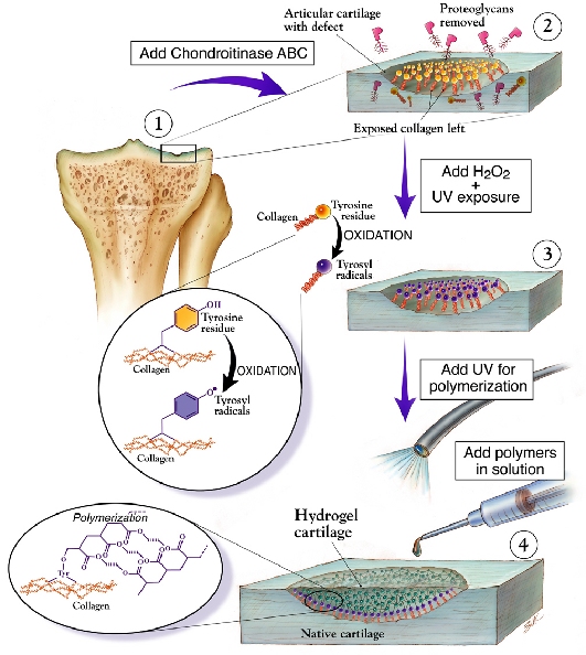
Hydrogels encapsulate cells and absorb a large volume of liquid for nutrient and waste transport. The poly(ethylene oxide)-based photopolymerizing polymers can incorporate, through physical interactions or covalent bonds, extracellular matrix derived gels and proteins. The composite hydrogel has a synthetic portion where physical properties can be easily manipulated and a biological portion where cell response and tissue development can be manipulated. Integration of a biomaterial with surrounding tissue is critical to long term survival and function. Particularly in the case of hard tissues such as cartilage in the musculoskeletal system, integration of an implant is difficult due to the dense nature of the extracellular matrix and large mechanical forces to which these tissues are often subjected. We have designed a method to direct covalent attachment of (meth)acrylated polymers to collagen proteins. Collagen is ubiquitous so that this method for implantation may be applied throughout. Covalent integration of hydrogel biomaterials significantly improves the mechanical integrity of the tissue-biomaterial interface in challenging applications such as cartilage. Photopolymerization is used as a method to encapsulate cells for drug delivery and tissue engineering applications in our minimally invasive implantation systems. Most tissues in the body are complex, containing numerous cell types organized in specific extracellular matrix architecture. We have created multilayered photopolymerizing hydrogels in order to encapsulate numerous cell types or drug delivery vehicles. The tissue is comprised of layers of chondrocytes which contribute to the unique developmental and mechanical properties of the tissue. Furthermore, osteochondral implants, containing bone and cartilage are often used clinically to treat joint defects. Local factors released by the various cell types influence engineered tissue development and properties. Using the appropriate cell type, we have created both multilayered cartilage and osteochondral constructs to create engineered tissue with enhanced biochemical and functional properties. A number of specialized hydrogels have also been optimized for specific cartilage regeneration applications. Proteoglycans are found throughout the body and serve numerous structural and functional roles. Chondroitin sulfate (CS) was modified to varying degrees with (meth)acrylate groups (CSMA) and polymerized to form a crosslinked hydrogel. Depending on the degree of modification, swelling, mechanical, and degradation properties of the CSMA hydrogels may be altered to fit a desired application. The CSMA gels are specifically degraded by chondroitinase and may be used as a diagnostic for the presence of chondroitinase, a degradative enzyme found in cartilage. Chondroitin sulfate is found naturally in other parts of the body including the intervertebral disc and may therefore be useful as a biomaterial in numerous applications. The diagram below demonstrates the process of using the photopolymerizable hydrogel for cartilage repair. You can click on the diagram to see a larger version.
Creation of new cartilage will use controlled delivery of biological signals mentioned above in addition to physical signals provided by a scaffold to design anisotropic, organized cartilage tissue. In addition, chondrocytes with varying morphology and gene expression may be organized during the encapsulation process. Tissue engineered cartilage with an organized structure similar to native tissue will have functionality comparable to native tissue and may potentially integrate more easily into the heterogenous host tissue where it is implanted. This three-dimensionality is expected to increase the speed and efficacy of tissue regeneration. Intellectual Property: Multiple patent applications are currently pending and reviewable upon the execution of a confidentiality agreement Elisseeff Receives Award MIT's Technology Review magazine recently recognized Jennifer Elisseeff, assistant professor of biomedical engineering at Johns Hopkins, as one of the World's Top 100 Young Innovators in technology and business. The Whitaker BME Institute With the nationally recognized Department of Biomedical Engineering as its core the Whitaker Biomedical Engineering Institute has been established as a vital link between the School of Medicine and the Whiting School of Engineering. The vision of the Institute is of an integrative research and education enterprise that will provide leadership in moving biomedical engineering to the forefront of biomedical science and practice. The Institute will be housed in Biomedical Engineering facilities in the School of Medicine and in Clark Hall, a new facility on the Homewood Campus. The Department currently has 17 primary faculty members, approximately 100 graduate students, and over 450 undergraduates. Ten new faculty members will be added to the Institute over the next six years, thanks to a Whitaker Foundation Leadership Award. The undergraduate program leading to the bachelor of science in engineering in biomedical engineering is now the largest program on the Homewood campus. Collaborators Dr.
Kam Leong, Ph.D.
Dr.
Kristi Anseth, Ph.D.
|
||
Useful
Link
|
| Technical
Contact Information:
Jennifer H. Elisseeff, Ph.D. Office Phone: 410.516.4915 |
Licensing
Contact Information: Phone:410-516-8300
|
|||
|
||||